


Structural features of the retina sensory part in experimental dysbiosis /hepatitis rats before and after taking blueberry paste
The research has been carried out at the histology laboratory of the Eye Diseases and Tissue Therapy Institute named after V.P.Filatov, Ukrainian Academy of Medical Sciences.
MATERIALS AND METHODS
Histomorphological research has been made on 12 rat eyes /female rats of Wistar line, aged 14 months/. The rats have been divided into three groups. The first group /4/ was the control one. In the second group /4/ dysbacteriosis+ hepatitis have been reproduced by introducing CCl and lincomycin. The animals of the third group /4/ with reproduced dysbacteriosis and hepatitis have been fed 2 g of blueberry paste daily together with their conventional feed.
After the experiment the eyes have been enucleated and fixed with 10 % formalin. The eyeballs have been put into paraffin. The paraffin sections have undergone hematoxylin-eosin and Van-Gieson staining.
All in all 12 items have been examined.
MORPHOLOGICAL FEATURES OF SENSORY RETINA WITH EXPERIMENTAL HEPATITIS+DYSBIOSIS
On modeling dysbacteriosis+hepatitis, sensory retina has been studied for pathological changes which have been found mainly in the ganglionic cells layer. These changes have not been very intensive and amounted to edematization combined with ganglionic cells axon garnetting. There have also been observed axon degeneration and hollows of various shapes and sizes. Vacuolar degeneration has been observed throughout the ganglionic cell cytoplasm (fig.1). Karyoplasm of the ganglionic cells nuclei has become clearer, granulosity typical of cytoplasm has disappeared. Areas with more intensive ganglionic cells vacuolar degeneration alternate with areas of less intensive degeneration or with those lacking degeneration at all. In cases of most intensive edematization and neuron degeneration the retina internal layers become netlike /fig. 2/.
The most intensive ganglionic cells vacuolar degeneration and retina internal layers edema have been observed in the spots corresponding to certain structural changes in retina blood vessels located mainly on the retina internal layers. Such changes have been found both in arterial and in venous vessels. It should be noted that the structure of various size arterial and venous vessels has been assessed as next to normal but there have been certain changes around them. These changes amount to small perivascular hoops composed of lymphocytes /fig.3/ and are accompanied by perivascular edema. In the spots where the retina changes are most intensive, there have been found slight eosinophilic homogenous masses, seemingly blood plasma effusions. This assumption is also confirmed by small-sized diapedesis haemorrhages observed both in the internal and in the middle retina layers /fig. 4/. Sometimes the retina internal layers have been found to
contain cavities made by erythrocytes, the histoarchitectonics of the external and internal nucleus layers being substantially destroyed.
No essential structural changes in the retina external layers have been observed.
All animals under the experiment have typically shown the signs of blood vessels thrombosis both in veins and in arterioles /fig. 4, 5/. Twice there have been found intensive destructive changes in blood vessels, their walls being homogenized and having no endothelial lining. The structures surrounding the vessels have been also degenerated /fig. 6, 7/, which is typically accompanied by retina internal layers dissection and accumulation of hemosiderin grains testifying to the previous haemorrhages.
SENSORY RETINA STRUTURAL CHANGES WITH THE USE OF BLUEBERRY PASTE
There have not been found any essential changes described above in the retina structure of the animals with reproduced disbacteriosis+hepatitis and on blueberry diet / fig. 8, 9/. The density of ganglionic cells has not been decreased. There have not been any structural changes in blood vessels.
CONCLUSION
Experimental disbacteriosis and hepatitis cause a number of structural changes in sensory retina. These changes are of dystrophy type and apparently are induced by toxic action of inflammatory reaction in retina as well as by circulatory disturbances.
The most intensive changes can be found in retina internal layers which indirectly testifies to the importance of circulatory disturbances to dystrophy development.
Despite a small number of animals under trials, we have discovered an explicit protective effect of blueberry preparation. The changes in the sensory retina have been reduced to just insignificant edema of ganglionic cells. There have been no disturbances in retina layers histoarchitectonics.
Fig. 1.Retina internal layers edema in rats with experimental disbacteriosis+hepatitis. Ganglionic cells polymorphism, internal plexiform layer edema, ganglionic cells vacuolar degeneration. No essential changes in other retina layers. Hematoxylin-eosin. X 600.
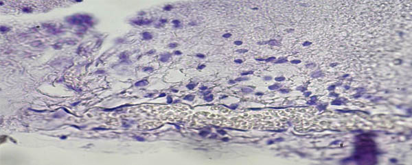
Fig. 2. Intensive edema of retina internal plexiform layer. Uneven distribution of polymorphous ganglionic cells. Net-like retina internal layers. Venule showing erythrocytes stasis. Hematoxylin-eosin. X 600.
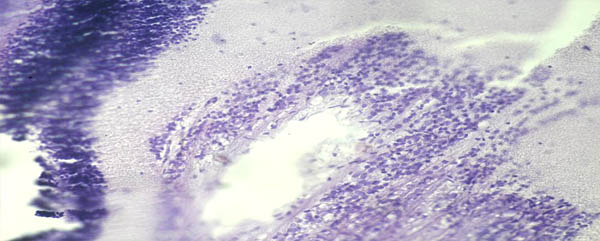
Fig. 3. Perivascular hoop consisting of pericytes and diffusely distributed lymphocytes. Endotheliocytes degeneration. Some endothelial cells are desquamated and provide for vein lumen. Hematoxilin-eosin. X 280.
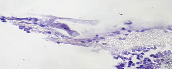
Fig. 4. Retina venule thrombosis accompanied by vessel endothelial lining destruction and retina internal and middle layers haemorrhages. Hematoxilin-eosin. X 600.
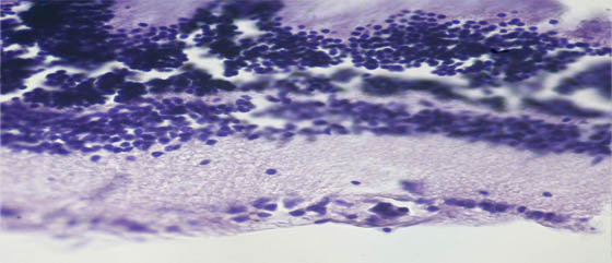
Fig. 5. Arterial vessel thrombosis in ganglionic cells layer. Homogenous thickening of capillary wall, edema and desquamation in ganglionic cells lumen. Hematoxilin-eosin. X 600.

Fig. 6. Changes in venous blood vessel wall in the retina internal plexiform layer. Vessel wall destruction. Retina internal layers detachment. Tissue imbibition with a small number of hemosiderin grains. Hematoxin-eosin. X 600.
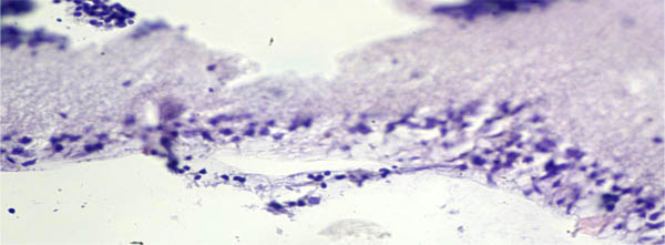
Fig. 7. Changes in venous blood vessel wall in retina ganglionic layer. Vessel wall homogenization, no endothelial lining. Retina internal layers detachment. Ganglionic cells vacuolar degeneration. Hematoxin-eosin. X 600.
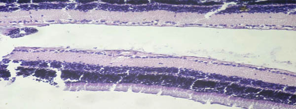
Fig. 8. Experimental animal retina on feeding blueberry paste. No structural changes in retina internal layers. Insignificant edema of ganglionic cells cytoplasm. Hematoxin-eosin. X 600.
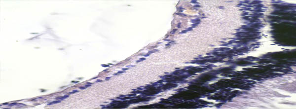
Fig. 9. Experimental animal retina on feeding blueberry paste. Insignificant edema of ganglionic cells cytoplasm. No changes in the arterial vessel. Hematoxin-eosin. X 600.
- TEK-PD cavitation-type dispersing pump
SIPE TEKMASH Institute offers dispersing pumps for making fine-dispersed food emulsions and suspensions, 10-25 m³/h capacity.
More... - Feedstock feeding equipment. Milk substitute.
The site contains information on fattening animals, on soy paste properties and soybean processing technology (animal feeding).
More... - Condensed milk line TEK-CML
Condensed milk production line
More...
- IndiaSASIL AGRO LLP
'Kshiti', 127/2, Rockel Lane,
Vakhar Bhag,
Sangli - 416416
MAHARASHTRA, India
Shreyans Shah
tel. +91-9011068877
e-mail: sasilexim@gmail.com - Colombia & LA
KAVITEC S.A.S.

Carrera 16 # 9-68
Dosquebradas, Risaralda, Colombia.?ngela Mar?a Sanz E
Gerente Comercial
email:
angela.sanz@kavitec.co
Tel?fono: 3306102
Cel: 3043289012 - Ukraine
© 1997-2022. All Rights Reserved.